Posts

Showing posts with the label microscope

Investigating the construction of a Bronze Age burial mound

Image
This post is many years in the making! Back in 2018 I was on an excavation at a site called Cronk Guckley on the Isle of Man with my colleague Prof. Chris Fowler, a brilliant theorist and Neolithic specialist, who is co-director of the Round Mounds project. This is a great project with some really exceptional finds, including this beautiful jet bead necklace. The excavation finished in 2022 and we are now completing all the post-excavation analysis. The project is investigating the nature and timing of Bronze Age ‘round mound’ construction and how funerary practices evolved, on the Isle of Man and surrounding islands. These structure are large earthern monuments that were constructed over the top of a burial. The jet necklace is an exciting piece of evidence, not just because of it's beauty, but because it gives us insights into the people - the jet has been sourced to Whitby, so is great evidence for connections between the Isle of Man and mainland Britain in the Bronze Age, an...

Phytolith mystery!

Image
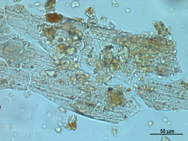
One of the nicest things about my job is hearing about the successes of past students. It makes me genuinely happy to see students with enthusiasm and passion for environmental archaeology go on to build successful careers. I taught my first postgraduate students during my time at Edinburgh back in  2013, and they are all doing so well. One recently finished a PhD and gained a great postdoctoral position, another is on track to finish their PhD very soon and has recently published their first paper, and another has a fantastic job as a research technician in a top environmental archaeology lab. I had an email from the latter recently regarding the identification of some unknown phytoliths. They appear to be generic grass long cells, but have odd striations that I have never seen before. The striations are all in the same direction - could this be an artefact of processing or is it surface decoration? I am not sure about surface decoration, it looks a bit too regular. has anyone see...

Micrograph of the Month: Inclusions in omnivore coprolite

Image
I am so happy that I've actually managed to get some microscope work done over the past couple of weeks. It feels like forever since I had the time to do any lab work, or spend time looking down the microscope. Even on my research leave, I have been so busy writing papers and grant applications that microscope time has taken a back seat. I am currently working on the 30 or so slides we collected as part of a NERC project at Paisley Caves in Oregon . This week I have been focusing on characterising the different types of faeces that are found in the sediments. There are lots of different types - rat and bat dung , ovicaprid type pellets, and of most interest of course, the potential human coprolites. I say potential, as we can't know for sure if they are human without conducting additional biomolecular analysis , but the pictures below show a likely human candidate. In any case we can say that it is omnivore coprolite, containing both digested bone fragments and plant tissues. T...

Micrograph of the Month: Mixed fuels

Image
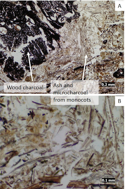
It's been too long since I've looked at any new thin section samples, but today I had the chance to look at some new stuff from Catalhoyuk . Here are some lovely mixed fuel deposits from an external area, showing A. wood charcoal (Quercus?), with microcharcoal and phytoliths from grasses and sedges. B. is a close up of the ash to try and show some of the phytoliths but it is hard to get a good photo in focus even though the slide is only 30 microns thick. Probably redeposited given the random orientation of the phytoliths.

Microfossil in a Micrograph of the Month

Image
It's been a while since I did a monthly microfossil or micrograph post, so I thought I would share this nice example of something combining both. This is a midden deposit from the early Neolithic site of Bonkulu in Turkey - the large greyish object in the middle is a very highly silcified conjoined phytolith from a reed stem/leaf, embedded in a matrix of mixed ashy debris. There is a resemblance to an earlier micrograph I posted from the Babylonian site tell Khaiber, which also contains large mutlicelled reed phytoliths. The deposits at Boncuklu are chock full of these large Phragmites reed phytoliths, likely to be from burning these substantial plants as fuel. The macrobotanical record from the site also contains large volumes of reed stem charcoal.

Micrograph of the Month: Roman floors

Image
I haven't posted a micrograph for ages, but this one is so pretty I just had to share it. I'm in the middle of doing an analysis of Roman occupation deposits for the Lufton Roman Villa project . I don't know too much about the wider context yet, but my initial observations of this particular sample suggests we are looking at an old floor surface. There are 3 distinct layers, and the uppermost layer is composed of a calcareous material, in which are embedded lots of tiny crushed up ceramic fragments. The middle layer is a sandy aggregate with the occasional bit of soil/clay and charcoal, and there is a very thin lowermost layer (thin because of the sample size, not sure yet how thick it was in the field) which has a lot of organic material including wood and grass charcoal, and possibly fungal spores. I will be writing more on this as the analysis progresses, but for now here is a nice image of one of the ceramic fragments (the orangey-red rectangular inclusion) embedded in...

Teaching archaeological sediment micromorphology part 2

This semester I've been teaching archaeological sediment micromorphology, as part of a third year module in geoarchaeology. I mentioned a few weeks ago how hard it was to teach a subject this complex in only 2 hours per week, and that in future I was thinking of removing it from Geoarchaeology and turning it into a stand alone module. I still haven't decided whether to do this, as I think it's important to have some teaching of it at UG level, but a module focusing entirely on micromorphology is probably more suitable for Masters level. Related to my frustrations, I made an offhand comment on Facebook that despite it being one of my specialist subjects, sediment micromorphology is very boring to teach. More than any other method I have studied, micromorphology is definitely the one filled with the most jargon. Don't get me wrong, I think jargon is important. We need the specialist language to describe the many complex features and processes that we see in thin sectio...

Thin sections related to pyrotechnology in Bronze Age Sicily

Image
The arrival of new thin sections is always exciting, but always daunting at the same time. Sediment micromorphology has to be one of the more challenging geoarchaeological techniques, simply because of the huge variety in the types of material you can encounter, and a deposit from one site never quite looks the same as one from another, even when they are related to similar activities. The samples from Case Bastione were collected to investigate formation processes and activities of a number of features in the Bronze Age settlement, including a large 'burnt' layer, and some strange pit deposits. The most striking thing about all the samples is the ubiquitous presence of these teeny tiny little creatures - my educated guess is that these are tiny foraminifera of some sort , present within limestone/chalk or another carbonate material (I need to do some research on Sicilian geology!). What is slightly confusing is the presence of a spherulite like appearance within the shells in...

Wolfson Archaeology Lab, Newcastle

Image
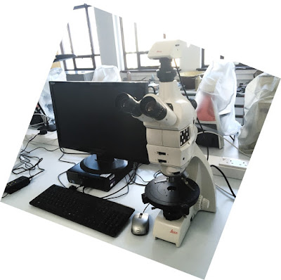
When I started at Newcastle one of the first major roles I undertook was taking over as Director of the Wolfson Archaeology Laboratory. This sounds rather grand, but in reality it mostly involves managing room bookings and making sure everyone has all the kit they need for practicals, research and teaching. Part of the job involves looking after a zooarchaeology reference collection, hence the post last month about attending the zooarchaeology  short course at Sheffield . I have also made various wonderful purchasing decisions, vastly expanding our suite of microscopes to include a range of Leica DM750P scopes for teaching, and dedicated scopes for research with even fancier specs including reflected light capabilities, image analysis software etc. The next stage will be to transform the side room, currently used mostly for storage, into a dedicated space for research. With a new lab based PhD and PDRA started in January, and possibly a Fellow later in 2017 (if all goes to plan), i...

Digitized thin section slides!

Image
I can't remember if I posted about this earlier in the year, but I was lucky enough to be awarded two student work experience placements as part of the  Newcastle NWE programme , where students complete flexible paid projects within the university. Two of my Environmental Archaeology students have been working for the past few months, digitizing my entire collection of thin section slides. At some point I hope to make these available online as an open access resource for teaching and research. They did a brilliant job! I've only just had a chance to go through all the scans, having been away on fieldwork, busy with exams, then graduation. Here is one of the scans of a thin section from medieval Riga, that I have been working on as part of the Ecology of Crusading project . Combined with the fact I have just moved the lovely Leica DM750P research microscope into my office (kindly purchased by History, Classics and Archaeology ), I can now get working on my mounting backlog of sa...

Micrograph: Layers within layers

Image
I love this image. It's another one from Catalhoyuk , a nice midden in the South Area (unit 17739). I published a paper on these deposits in Antiquity which included this image , so I'll let you read the paper to find out more about this area and its significance. Here I wanted to show a close up of this image and the beautiful but daunting complexity of archaeological deposits under the microscope. What we are looking at is a tiny fragment of wall plaster mixed in with ashy debris and charcoal. A few years ago I did a post about these plaster deposits, as they are found within buildings at Catalhoyuk , on floors and walls. By counting the layers we can see the frequency with which the inhabitants were re-plastering and 'repainting' their houses - regular cycles of maintenance on an annual and seasonal basis. This layer in the midden shows a fragment which has fallen off a wall, and somehow made its way into the midden, probably through sweeping and dumping of debris, ...

Micrograph: Partially burnt sediment with plant voids

Image
I've been preparing for teaching a micromorphology lab class tomorrow, on the topic of architectural materials. Having recently dug out my old laptop ( to look at old job applications... ), I came across my huge archive of micromorphology images. I really need to figure a way of making these available online. I have so many and they are such fantastic references. I found this nice example of a partially burnt aggregate; it is perfect for showing how pseudomorphic plant voids appear in a plant-tempered clay. On the right hand side of the image, the plant temper (grasses) has been burnt, and so has preserved in the form of microcharcoal. On the left hand side, where the aggregate escaped the burning, the plants have totally decayed, leaving only the voids in the shape of the grasses. It also shows how the colour of the sediment change as they are heated, becoming a distinctly darker brown. This sample is from the Deep Sounding midden at Catalhoyuk, some of the earliest deposits that...

Make your own archaeological poop

Image
Two posts in one day?? Yes, deadlines are looming so the blog posting is on fire. I thought I would do a little update on some non-academic stuff I've been doing recently. Around this time last year I ran an Indiegogo fundraiser . The main reason was that I no longer had an academic job, and therefore no longer had access to a microscope to continue my research that I had started at the Ness of Brodgar in Orkney. I was unsure about whether to do crowdfunding, it is really hard asking individuals for money, compared to asking research councils for millions (that's harder in a different way). But I couldn't think of any other options and desperately wanted to continue the research I was doing. The campaign was reasonably successful; I didn't raise the total, but I did get enough to buy a halfway decent microscope and to cover the costs of taking it up to Orkney for the next field season, to do a 'field lab' where students and visitors could watch the analysis in p...

Microfossil of the Month: Discus diatoms

Image
Almost forgot to post a microfossil for November! That would be sad as I've managed to do the monthly micrograph then microfossil posts every month for almost two years now. I've just been so busy with teaching, and the lack of microscope camera makes taking pictures a bit more of an effort than it used to be. Good news is that we do have a new set of microscopes and cameras for the archaeology lab here at Newcastle, hurray! The lab however is located in a different building to my office, so photograph acquisition still requires a bit more planning than it used to. As it is almost the end of 2015 (yikes), I am thinking of a new monthly blog feature, perhaps a thin section of the month, where I show you exciting pictures of whole thin section slides! But for now, here is something very pretty. Like the sponge spicules I posted earlier in the year , these little creatures are not my specific area of expertise, but they occasionally show up in my phytolith slides. They are littl...

Microfossil of the Month: Which Wood?

Image
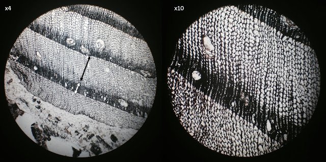
Something a little bit different for October. This fits in nicely with what I've been up to this week, preparing teaching materials on wood charcoal analysis in archaeology. Although I am not technically a wood specialist, I am wondering if I should pick up a new skill set, as it would actually be very useful as a thin section micromorphologist. This image shows a cross section through an unidentified fragment of wood charcoal in a thin section of sediments from the tower at Cesis Castle, Latvia . There are layers within the sediment that are full of tiny wood charcoal fragments, and actually we see bits of wood a lot when looking at thin sections of ash samples, unsurprisingly. You can see the annual rings quite clearly in this fragment, and it can be identified as a soft wood species (coniferous) due to the lack of pores. Soft woods are a bit more difficult to identify than hard woods (from deciduous trees), as their structure is more simple and less distinctive. In this example ...

Microfossil of the Month - Globigerina Ooze

Image
A bit of a geoscience themed iPhone micrograph for September (I am getting quite good at this iPhone down the eyepiece photo taking thing). Yes, there really is a sediment called ooze... Globigerina  ooze! Archaeologists are probably aware that much of our global scale climate reconstructions come from the isotopic analysis of deep sea sediment cores, and  Globigerina ooze is one of these sediments. Huge areas of the ocean floor are covered in this stuff, which consists largely of the shells of various foraminifera, of which the species Globigerina bulloides is the most common. The shells of these little creatures are composed of calcium carbonate (CaCO 3 ) and it is the oxygen component of this that is used for oxygen isotope analysis in the  reconstruction of  temperature in the past. The name  Globigerina  ooze was first used to describe the sediment during planning and construction of the earliest transatlantic telegraph cables. They are ve...

Ochre in Orkney

Image
Oh the excitement! Today I received a parcel from Earthslides , with 20 new thin section samples from the Ness of Brodgar . These samples were collected in 2014 and are mostly from the Trench T midden. Although I don't yet have the kit in place to do a proper analysis, I couldn't resist having a quick peek with my old Swift scope. The level of complexity is daunting - so may fine layers of stratigraphy hidden within layers that appear relatively homogeneous in the field. But already I can see something interesting things - layers of burnt peat,  a very large amount of burnt bone, and even a tiny fragment of red ochre pigment, about 2mm in diameter. This pigment has been found in various contexts in Orkney (and elsewhere around the world), for example Gordon Childe at Skara Brae found containers of pigment he interpreted as 'paint pots '.  At the Ness it is thought that the pigment was ground down and used as a paint for the stone walls in buildings. Behold my attempts a...

Microfossil of the Month: Fragile phytoliths

Image
I promise brand new images will be coming soon, but for now here is a micrograph from my old files, this time from my work with the Central Zagros Archaeological Project back in 2010, and is from ashy deposits in an external area at the Neolithic site of Sheik e Abad in Iran. This is a great example of a conjoined phytolith that is not particularly well silicified and/or has suffered erosion. The pattern of the cells is not very distinct, and quite 'faint', though you can just about make out the wave pattern of the long cells in places. For comparison, see this example of well-silicifed wheat phytoliths from Catalhoyuk, and this reference specimen of Setaria italica , both of which have very distinctive and well defined cell morphologies. The reason I chose this micrograph is that it is a very good example of how fragile phytoliths are. Despite being composed of silica, which is pretty resistant to decay, phytoliths are physically quite fragile. Think of it like glass - it i...

Microfossil of the Month: Setaria italica tissue phytoliths

Image
Last month I thought that I would be unable to post any new micrographs for a while, however with a stroke of luck, and digging out of an old hard drive, I came across all the files from my PhD thesis! A stark reminder of the importance of proper archiving of digital image files, I must have hundreds of images that have not been catalogued properly...I'll add that to the to do list! Here we have a micrograph of a reference specimen of Setaria italica, more commonly known as foxtail millet. In this image you can see that this is a very well silicified bit of plant tissue, with all the individual cells being clearly defined. This is a leaf fragment and you can see the spikey hair phytoliths, which are also called trichomes. the little 'dumb-bell' shaped short cells are known as bilobes or bilobate cells. These bilobes are typically found in plants with C4 photosynthesis, and can give an indication of the broad type of environment. C4 refers to the biochemical mechanism that ...

Castles and Coprolites - now on video!

Image
Just taking this opportunity to share a video I recently posted on my YouTube page, from my talk at the Archaeological Research in Progress conference 2015, organised by the Society of Antiquaries of Scotland . Any feedback and comments on this would be much appreciated, as it is the first 'full length' talk I have done that has been recorded (by the talented Open Access Archaeology ). I now have a grand total of 3 videos posted on YouTube, and it got me thinking about the idea of doing video-blogging. Is this something that people would find interesting? I was thinking of doing short videos that summarise different aspects of my work (and related research areas), and including PowerPoints that I have previously used in my teaching - this could either be at a very introductory level, or a little more advanced. What sorts of topics would people like to see? Also taking this opportunity to share my IndieGoGo campaign again, which is now half way through and on 21% - thank yo...