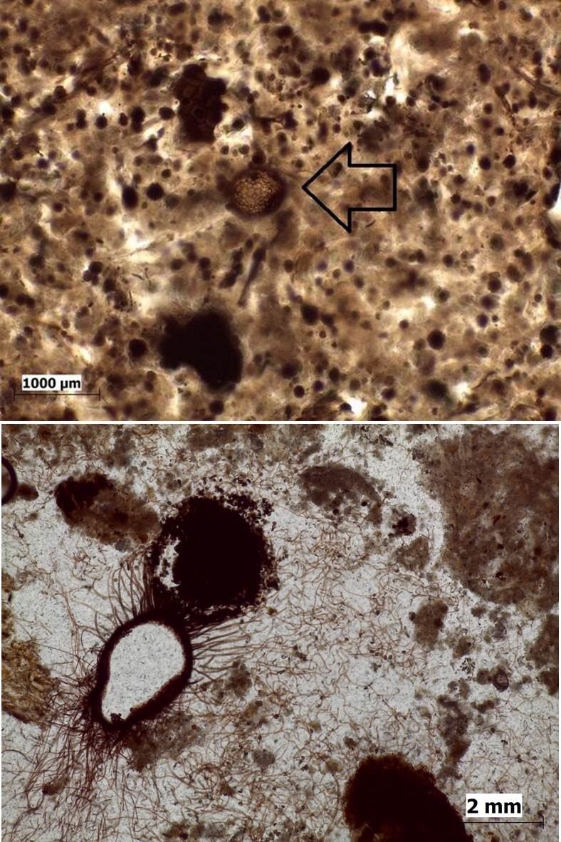
Followers of my Twitter account may recognise these little creatures! I posted the pics separately a while ago to see if anyone could help with identification. So far no response, if anyone has any clues or reference suggestions do let me know. Both are from medieval floor deposits that have undergone significant post-depositional bioturbation. The lower image is one of my favourites. The hyphae are like little tentacles that spread all the way through the sediment. That is the little string like projections that you can see extending from the sporongium. Which is the spherical bit containing all the little spores, and in this particular view is nice and ripe with little spores bursting forth. It is quite creepy to come across all of a sudden when you are looking down a microscope and not expecting it! The one at the bottom has a clear area in the middle, because the top has been abraded away during the thin section preparation process. Basically we are looking at an 'aerial' view of the 'heads' if the fungus, which was standing up at a thickness of greater than 30 microns. In the upper image the sporongium is still quite pale and still in the process of growing. Or at least it was until it was set in resin and turned into a slide.


Comments
Post a Comment