Posts

Showing posts with the label phytoliths

X-rays and Barley Mutants!

Image
Blog readers may be familiar with the botanical terms monocots and dicots. Dicots include many woody species, and monocots include important cereal crops such as wheat and barley (i.e. grasses). These are basics of plant biology that I teach my students in environmental archaeology. Monocots such as cereals of course are central in archaeological research around the origins of agriculture and domestication, whilst dicots feature in studies of ancient fuel use.  One of the major differences between monocots and dicots is the organisation of the stem. Dicot stems, or cambium, have radial growth, that forms the distinct rings that we use for dendrochronology. This growth is controlled by the interaction of two proteins, TDIF and PXY. Monocots do not have a cambium, but they do have the genes for TDIF and PXY. What then, is the function of these genes in monocots, if not to produce cambium? This question is the basis of a major BBSRC funded project I've been involved in for t...

Human-environment interactions in the Hadrian's Wall Landscape

Image
You wait a whole year for a blog post, then two come along at once etc. Following the existential musings of the previous post, I figured I'd better post some fun geoarchaeological stuff. I have been working with a number of colleagues on various projects for the past couple of years, that are loosely related, and fall under the general banner of Human-environment interactions in the Hadrian's Wall Landscape. Regular blog readers will remember in 2021 I talked about the new  excavations at Birdoswald . These have been progressing nicely, and we have uncovered all sorts of amazing finds and environmental material. I have also been working with Dr Eline Van Asperen, and Masters student Damian Rudge, looking at long term changes in the environment, and whether these relate to Roman or earlier activities such as lead mining. In 2022 we were awarded a small grant from the Royal Archaeological Institute  to collect some peat cores with the aid of local volunteer groups. Eline a...

Phytolith mystery!

Image
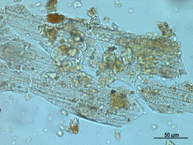
One of the nicest things about my job is hearing about the successes of past students. It makes me genuinely happy to see students with enthusiasm and passion for environmental archaeology go on to build successful careers. I taught my first postgraduate students during my time at Edinburgh back in  2013, and they are all doing so well. One recently finished a PhD and gained a great postdoctoral position, another is on track to finish their PhD very soon and has recently published their first paper, and another has a fantastic job as a research technician in a top environmental archaeology lab. I had an email from the latter recently regarding the identification of some unknown phytoliths. They appear to be generic grass long cells, but have odd striations that I have never seen before. The striations are all in the same direction - could this be an artefact of processing or is it surface decoration? I am not sure about surface decoration, it looks a bit too regular. has anyone see...

Microfossil in a Micrograph of the Month

Image
It's been a while since I did a monthly microfossil or micrograph post, so I thought I would share this nice example of something combining both. This is a midden deposit from the early Neolithic site of Bonkulu in Turkey - the large greyish object in the middle is a very highly silcified conjoined phytolith from a reed stem/leaf, embedded in a matrix of mixed ashy debris. There is a resemblance to an earlier micrograph I posted from the Babylonian site tell Khaiber, which also contains large mutlicelled reed phytoliths. The deposits at Boncuklu are chock full of these large Phragmites reed phytoliths, likely to be from burning these substantial plants as fuel. The macrobotanical record from the site also contains large volumes of reed stem charcoal.

Did the people of Çatalhöyük build boats?

Image
Two posts crossed my Twitter feed last week that got me thinking. Both are related to experimental archaeology and use of wetland resources. If you are a regular reader of my blog (or indeed my academic papers) you will have noticed that I talk a lot about the environment of Çatalhöyük, and how this may have influenced human activity in the Neolithic. You will also notice that I have criticised some of the early interpretations which, to me, present a simplistic interpretation of evidence that doesn’t consider the complexity of the environment, or people. Çatalhöyük was situated in a wetland environment, and there have been debates over the influence this had on agriculture. Where were people growing their crops if the local area was not suitable for agriculture?  One theory suggests people travelled to fields that were located many km away from the site, which in turn has implications for social organisation. This always bothered me, firstly because of the limitations of the ...

The details of giant daisies

Image
Today is the end of the Semester, and I'm definitely ready for a break after an extremely busy few months. Lots of great news this year, with two successful grant applications - the NERC project which I've been posting about, and a new Wellcome seed award which will be starting next year, developing further work at Catalhoyuk in Turkey. I have also had my own assignment deadline this week, for the NERC training course that I am going on in January . Everyone in the group has been assigned a plant family, and we had to do a bit of research on the genera and species that are found in Belize, and write a short information sheet on key characteristics. I think the expedition leader took pity on me as one of the participants without a background in botany, and I was assigned the Asteraecae (Compositae), which happen to be one of the easier families to identify. Or at least, it is one I am familiar with - daisies and sunflowers! Some of the terminology was familiar, ringing bells fro...

Training in Belize!

Image
2016 - the year where the exciting news just keeps on coming! I found out recently that I've been selected to take part in a NERC training course, Fieldwork Skills in the Tropics, run by the Royal Botanic Gardens Edinburgh , as part of their MSc in Biodiversity and Taxonomy of Plants . The course runs over two weeks next year, in Belize! It's been over a decade since I first left the UK on my first fieldwork abroad, but the thought of travelling somewhere new is still as exciting now as it was then. Probably even more so as I have some idea what I'm doing now. This course covers vegetation survey, monitoring and plant identification, with some GIS. Part of the application process involved a statement of why you would like to attend, with priority given to those with NERC funding, ECRs and students. Whilst I now work as an archaeologist, my background is in geosciences/geography, and I've always maintained that perspective in my research and teaching. My long term aim is...

Micrograph: Layers within layers

Image
I love this image. It's another one from Catalhoyuk , a nice midden in the South Area (unit 17739). I published a paper on these deposits in Antiquity which included this image , so I'll let you read the paper to find out more about this area and its significance. Here I wanted to show a close up of this image and the beautiful but daunting complexity of archaeological deposits under the microscope. What we are looking at is a tiny fragment of wall plaster mixed in with ashy debris and charcoal. A few years ago I did a post about these plaster deposits, as they are found within buildings at Catalhoyuk , on floors and walls. By counting the layers we can see the frequency with which the inhabitants were re-plastering and 'repainting' their houses - regular cycles of maintenance on an annual and seasonal basis. This layer in the midden shows a fragment which has fallen off a wall, and somehow made its way into the midden, probably through sweeping and dumping of debris, ...

Micrograph: Unusual archaeobotany

Image
As a micromorphologist something that you begin to notice quite quickly is that plant remains come in a huge variety of forms. Whilst at the macroscale we usually think about charred remains - wood charcoal, burnt seeds and grains - at the microscale there is a whole world of other plant remains. Phytoliths and other microfossils are something I talk about quite a lot in this blog, and these are becoming a standard form of 'plant evidence' alongside plant macros. More unusual are pseudomorphic voids, plant remains that are no longer there! I have talked about these a lot in previous posts , basically they are 'impressions' of plants that have since decayed. The micrographs below show something a bit in between. The plants haven't fully decayed, as they were partially desiccated. The orange colour you can see is where the organic matter from the plant has stained the calcareous aggregate (probably some sort of architectural material). In the voids you can see remains...

AEA conference 2015 - some thoughts on taphonomy, equifinality, and multi-proxy approaches

This weekend I went to my first AEA conference . As a student, then as a fixed term post-doc, it is difficult to fund conference attendance, and also to find the time when there are so many conferences to choose from. Having done a few years of the big conferences ( SAA and EAA ) to maximise audience and networking opportunities, I’ve decided to spend some time at the smaller ones, where I can focus on my specific interests. Overall it has been an enjoyable weekend, and it was great to be back in York and catch up with old friends and colleagues, including Matthew Collins , who I can’t thank enough for writing me many references over these past few years (I bet he’s relieved that I finally got a job so he can stop writing them!). There are three ‘themes’ that stood out for me at the conference. The first was the study of taphonomy, and how wildly different this is between different techniques in environmental archaeology. The second was the recognition of the advantages of inte...

Microfossil of the Month: Fragile phytoliths

Image
I promise brand new images will be coming soon, but for now here is a micrograph from my old files, this time from my work with the Central Zagros Archaeological Project back in 2010, and is from ashy deposits in an external area at the Neolithic site of Sheik e Abad in Iran. This is a great example of a conjoined phytolith that is not particularly well silicified and/or has suffered erosion. The pattern of the cells is not very distinct, and quite 'faint', though you can just about make out the wave pattern of the long cells in places. For comparison, see this example of well-silicifed wheat phytoliths from Catalhoyuk, and this reference specimen of Setaria italica , both of which have very distinctive and well defined cell morphologies. The reason I chose this micrograph is that it is a very good example of how fragile phytoliths are. Despite being composed of silica, which is pretty resistant to decay, phytoliths are physically quite fragile. Think of it like glass - it i...

Microfossil of the Month: Setaria italica tissue phytoliths

Image
Last month I thought that I would be unable to post any new micrographs for a while, however with a stroke of luck, and digging out of an old hard drive, I came across all the files from my PhD thesis! A stark reminder of the importance of proper archiving of digital image files, I must have hundreds of images that have not been catalogued properly...I'll add that to the to do list! Here we have a micrograph of a reference specimen of Setaria italica, more commonly known as foxtail millet. In this image you can see that this is a very well silicified bit of plant tissue, with all the individual cells being clearly defined. This is a leaf fragment and you can see the spikey hair phytoliths, which are also called trichomes. the little 'dumb-bell' shaped short cells are known as bilobes or bilobate cells. These bilobes are typically found in plants with C4 photosynthesis, and can give an indication of the broad type of environment. C4 refers to the biochemical mechanism that ...

Microfossil of the Month: Multi-celled Sedges

Image
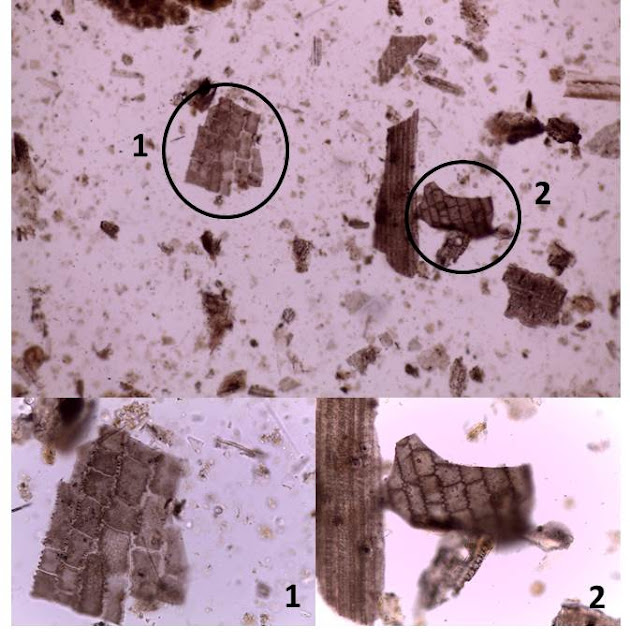
Yikes, I can't believe it's already June, and time for a new microfossil! This month we have firstly a general view of a phytolith slide from the site of Boncuklu in Turkey , at x100. This just gives you a taste of how chock full of microfossils these slides are! I have highlighted two particular phytoliths here shown at x400, both are conjoined phytoliths from sedge. Sedges, or Cyperaceae, are monocots which are similar to reeds, and are associated with wetlands. Though significantly, it should be noted that they can be found in other types of environment as well. At Boncuklu we know from other environmental work that a local wetland habitat was quite likely, and we also see a lot of reed phytoliths in these samples. The blocky square pattern is typical of sedge phytoliths, though it is difficult to say anything about which species they might be from. This sample is from an ashy layer in a midden deposit, and it could be that the sedges were burnt alongside reeds, either delib...

Microfossil of the Month: Wheat husk phytoliths

Image
This month's microfossil is a classic, at least within near eastern archaeology. The beautiful little structure you are looking at here is a phytolith from the husk of wheat. A huge area of research in phytolith studies is focused on cereals, and whether cereal phytoliths can be used to identify the genus or even species of cereals, and whether we can distinguish cultivated cereals from wild grasses. This obviously has very significant potentials in studying the origins and development of agriculture. This particular phytolith is from the middle Neolithic levels at Catalhoyuk , Turkey. At this point we have definitive evidence for agriculture from other lines of evidence, such as charred cereal grain stores within buildings. This phytolith was recovered from a midden, and is interesting because of its size. There is a positive correlation between the size of conjoined phytoliths and the availability of water during the growth of the plant. you can see that this phytolith is very la...

Microfossil of the Month: Awn phytoliths

Image
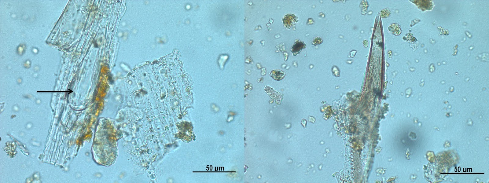
These micrographs show grass awn phytoliths, on the left the spiky hair part is still attached to the rest of the awn, the one on the right shows a hair that has become detached. Awns are hair or bristle type structure which are particularly common on grasses. In some species, such as emmer wheat, the awns contribute to seed dispersal, by hooking into the soil. Changes in humidity cause the awns to expand and contract, acting like a ratchet to propel the seed into the ground. Not to be confused with trichomes, which are another 'hair-like' structure, but a much finer growth of hairs on the surface of the epidermis, and can also produce distinctive phytoliths. Both of these examples are from the same sample, extracted from ashy deposits in an external area at Sheik-e Abad, an early pre-pottery Neolithic site in Iran. The site was excavated as part of the Central Zagros Archaeological Project . I worked there for 2 months after submitting my PhD in 2008, one of the most fascinat...

Microfossil of the Month: Plant Stomata

Image
For 2015 I have decided to replace my Micrograph of the Month feature with Microfossil of the Month. I will likely return to the thin section micrographs at some point, but as I am going to be working on phytolith and ash samples for most of this year, I figured it would make sense to feature some of these on the blog! Who knows how long I will be able to keep it up - from past experience of the sites I am working on, I may be limited in the range of examples that I will find! As a refresher (and for those of you new to the blog), microfossils are any fossil remains that are too small to see easily with the naked eye, but can be identified under the microscope. I work on plant microfossils, specifically silica phytoliths, which are 3D 'impressions' of plant cells and tissue. When the plant is alive, it takes up silica from the ground, which is then deposited inside and between the cells, forming 3D replicas of the cells that preserve when the organic part of the plant decays. T...

Goats and reeds in the Neolithic

Image
Bulliform cells in modern Zea mays tissue (source: Visuals Unlimited, Inc) Fellow animal dung enthusiast Sarah Elliot at the University of Bournemouth shared this fab little article describing the use of 'Eco goats' to manage invasive plants in the US . It is especially interesting that they note the use of goats as a pesticide-free way to manage Phragmites reeds. These reeds have distinctive phytoliths which archaeologists call 'keystone' bulliforms, which are relatively large single cell phytoliths that are named as they are said to resemble a Roman keystone arch in cross section.  In transverse section they look a bit sausage shaped, and within a plant these cells are 'stacked' next to each other as part of the upper epidermis (the plant 'skin'), and are related to water storage. If you're a botanist you might also call these motor cells, because during times of water stress, they shrink and cause leaves to fold or curl up. Phragmites phytoli...

Micrograph of the Month: Layers of reeds

Image
A little bit late this month as I've recently moved office and only just had my microscope camera software installed on my new PC. Incidentally this is also the reason I haven't added scale bars to these images, as I haven't had the chance to calibrate the magnification for the software (you have to tell the computer what the magnifications are by taking measurements of known lengths on a micrometer). The image to the left is at x10 magnification, the one on the right is x20. Here we are looking at some ashy deposits from the Babylonian city of Tell Khaiber, Iraq (being excavated as part of the Ur region project ). These are absolutely full of plant phytoliths and grass derived microcharcoal. The structure of these conjoined cell phytoliths is beautifully preserved. I have highlighted the multiple layers of plant tissue that are likely to be from reeds (a bundle of stems?). The stacked bulliform phytoliths are typical of reeds, and are often seen as individual cells. Here w...

Ethnoarchaeology - animal dung in Iraqi Kurdistan

Image
I've just been notified of the publication of this great new paper via Google Scholar citations, as it cites my paper on lipid analysis of coprolites . That's not the only reason I'm writing about it mind you - anything dung related gets my attention, and this is one of the first papers to come out of the Ce ntral Zagros Archaeological Project , which I was involved with a number of years ago. The CZAP project aims to understand the origin and process of animal domestication and agriculture in the Central Zagros region of Iran and Iraq, and the team have also conducted ethnographic studies in modern villages in the area, in order to help interpret the archaeology. This paper by Elliot et al reports on a mix of ethnographic studies and scientific analysis of modern dung and plants. The authors look at the dung spherulites in modern samples of sheep, goat and cows - those little spherical particles that form in the guts of animals . Despite being used frequently to support t...

Microstratigraphy and micro CT scanning

I came across these two studies in Journal of Archaeological Science recently, through Google citation alerts . It's always satisfying when my papers are cited, as it shows that there are people out there actually reading them and finding them useful! It's especially satisfying when the citing papers are as exciting as these! Both are by the same team, with lead author Hans Huisman who you may remember gave me some very useful advice through my Micrograph of the Month feature on some of my thin sections. The first paper is " A question of scales: studying Neolithic subsistence using micro CT scanning of midden deposits " Here Husiman et al. apply micro CT scanning to thin section analysis. CT scan, or CAT scan, stands for computerised tomography, which uses a series of X rays to produce 3D images of components that are enclosed within a matrix. This is the same technique that is used in medical applications to look inside the body, but at a much smaller scale, hen...