Posts

Showing posts with the label taphonomy

Adventures in medium sized mammal bone preparation, part 2

Image
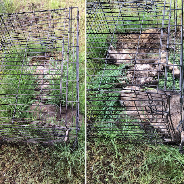
Well, I arrived in Oregon a couple of days ago, trying (without auccess) to get over jet lag before starting firldwork fot the NERC project tomorrow. Before I start with the stream of NERC related posts, a little update on my taphonomic experiment that I posted about back in November . Readers may recall that we came across some recently deceased racoons plus a hawk on the side of the road, and I decided they would make a fine addition to my animal bone reference collection. I set them up in a wire cage to be left to the elements, thinking that when I came back 6 months later they would be in the advanced stages of decay, perhaps even ready to extract and clean up the bones. Nope. 6 months sounds like plenty of time for two medium sized mammals plus one hawk to decay, but I didn't account for the fact that when I deposited them back in November, winter was coming, and they have been buried under two feet of snow for the best part of those 6 months! So, they pretty much look like th...

AEA conference 2015 - some thoughts on taphonomy, equifinality, and multi-proxy approaches

This weekend I went to my first AEA conference . As a student, then as a fixed term post-doc, it is difficult to fund conference attendance, and also to find the time when there are so many conferences to choose from. Having done a few years of the big conferences ( SAA and EAA ) to maximise audience and networking opportunities, I’ve decided to spend some time at the smaller ones, where I can focus on my specific interests. Overall it has been an enjoyable weekend, and it was great to be back in York and catch up with old friends and colleagues, including Matthew Collins , who I can’t thank enough for writing me many references over these past few years (I bet he’s relieved that I finally got a job so he can stop writing them!). There are three ‘themes’ that stood out for me at the conference. The first was the study of taphonomy, and how wildly different this is between different techniques in environmental archaeology. The second was the recognition of the advantages of inte...

Microfossil of the Month: Fragile phytoliths

Image
I promise brand new images will be coming soon, but for now here is a micrograph from my old files, this time from my work with the Central Zagros Archaeological Project back in 2010, and is from ashy deposits in an external area at the Neolithic site of Sheik e Abad in Iran. This is a great example of a conjoined phytolith that is not particularly well silicified and/or has suffered erosion. The pattern of the cells is not very distinct, and quite 'faint', though you can just about make out the wave pattern of the long cells in places. For comparison, see this example of well-silicifed wheat phytoliths from Catalhoyuk, and this reference specimen of Setaria italica , both of which have very distinctive and well defined cell morphologies. The reason I chose this micrograph is that it is a very good example of how fragile phytoliths are. Despite being composed of silica, which is pretty resistant to decay, phytoliths are physically quite fragile. Think of it like glass - it i...

Micrograph of the Month: Bits of Bones

Image
For the past few weeks I have been revisiting some old slides from the TP excavation area at Catalhoyuk . I collected these samples  in 2004, and they were some of the first that I worked on for my PhD. At the time I found them a bit disappointing, as there is a lot of bioturbation and erosion  going on in this part of the site, which means that trying to reconstruct activities using microstratigraphic analysis is difficult. The TP area is located very close to the surface of the mound, so despite the fact that these are the youngest deposits, the preservation is nowhere near as good as the earlier, deeply buried deposits. In the end I focused more on the South and 4040 areas, with the TP samples being used as a brief comparison of how different the taphonomic processes are in different parts of the site. Which brings me on to this month's photos! I was contacted by Kamilla  PawÅ‚owska who is conducting zooarchaeological analysis in the TP area, and is...

Micrograph of the Month: Dissolution of Spherulites

Image
This post follows on from the Manganese Micrograph Mystery I posted a while ago. Blog followers will remember I posted some images of layered ruminant dung which had a distinctive black 'speckled' appearance, a bit similar but not quite the same as Mn staining. Thanks to my readers the mystery was solved through the kind contributions of Hans Huisman. Huisman is a geoarchaeologist with expertise in degradation and preservation of archaeological materials. He suggests that manganese would produce more dendritic like patterns, and that instead this appears to be iron sulphide staining. Iron sulphides include minerals such as pyrite, which can oxidise to rust . This type of staining can be recognised in OIL as having a 'metallic' lustre, or smaller particles, such as we have here, can be examined using reflectance microscopy at a high magnification. So I'll be doing that asap. For now I wanted to share this image showing the possible impact this staining has had on ...

Micrograph of the Month: Modern roots

Image
In your sediments, post-depositionally disturbing your stratigraphy. Here we have examples of roots from modern plants that have grown into the archaeological deposits. They are easily distinguishable from archaeological plant remains as the tissue is fresh, organic and well preserved. Photo A shows a root that has grown into soft, ashy, compact midden at Boncuklu, Turkey. You can see clearly the void that has been created from the root's growth, which is the same shape as the root itself (3). We can also see a fragment of shell (1) and some small crushed bone fragments (2) embedded in the deposits. Photos B and C show close ups of modern root from Cesis Hillfort, Latvia. In B you can see where the root is divided into two rootlets whilst C shows the cell structure very clearly. Each of those 'jigsaw' shapes is an individual cell - it is cells like this that become infilled with silica during the growth of the plant, to produce phytoliths. This example of 'jigsaw' s...